Severe Anemia Case Study: Lab Interpretation Guide for Accurate Diagnosis

Introduction to Severe Anemia Case Study (Step-by-Step Lab Interpretation)

The Severe Anemia Case Study (Step-by-Step Lab Interpretation) is designed to guide healthcare students and professionals through the logical process of diagnosing severe anemia using laboratory data. Severe anemia isn’t just “low hemoglobin.” It’s a potentially life-threatening condition that demands rapid interpretation and precise clinical reasoning.

In this article, we’ll break down a real-world case in a structured, practical way. We’ll examine laboratory values, interpret them step by step, explore differential diagnoses, and determine the most likely cause—just like you would in clinical practice.

Let’s dive in.

Understanding Severe Anemia: Definition and Clinical Significance

What Is Anemia?

Anemia is a condition in which the hemoglobin level is lower than normal for age and sex. Hemoglobin carries oxygen to tissues, so when levels drop, organs don’t receive enough oxygen.

Normal hemoglobin levels:

  • Men: 13.5–17.5 g/dL

  • Women: 12.0–15.5 g/dL

When Does Anemia Become Severe?

Anemia is considered severe when:

  • Hemoglobin < 7 g/dL (critical threshold)

  • Patient shows symptoms of hypoxia or cardiovascular instability

Severe anemia can cause:

  • Fatigue

  • Shortness of breath

  • Chest pain

  • Tachycardia

  • Dizziness or syncope

Now let’s examine a clinical case.

Case Presentation: Patient History and Symptoms

Demographic Information

  • 38-year-old female

  • History of heavy menstrual bleeding

  • Vegetarian diet

Presenting Complaints

  • Progressive fatigue for 3 months

  • Shortness of breath on exertion

  • Palpitations

  • Headache

Physical Examination Findings

  • Pale conjunctiva

  • Tachycardia (110 bpm)

  • Mild systolic murmur

  • No lymphadenopathy

These findings suggest chronic anemia.

Step 1: Complete Blood Count (CBC) Interpretation

ParameterResultNormal Range
Hemoglobin6.2 g/dL12–15 g/dL
Hematocrit19%36–46%
MCV68 fL80–100 fL
MCHLowNormal
RDWHigh11–15%

Hemoglobin and Hematocrit Analysis

The hemoglobin of 6.2 g/dL confirms severe anemia. The low hematocrit supports this finding.

Red Blood Cell Indices (MCV, MCH, MCHC)

MCV is 68 fL → Microcytic anemia
Low MCH → Hypochromic cells

Microcytic, hypochromic anemia strongly suggests:

  • Iron deficiency anemia

  • Thalassemia

  • Anemia of chronic disease

Red Cell Distribution Width (RDW)

High RDW indicates variation in red blood cell size (anisocytosis).
Iron deficiency anemia typically has high RDW.

This narrows the differential..

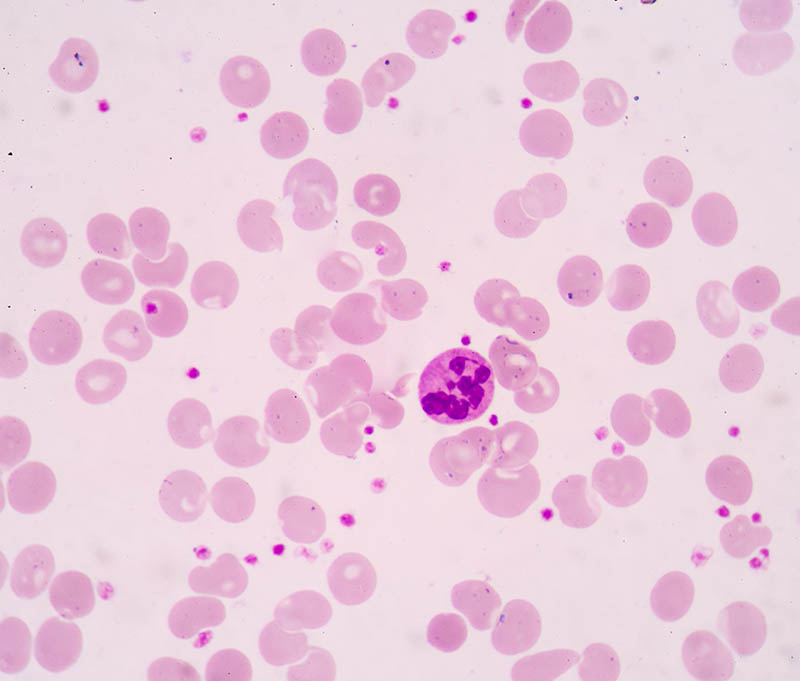
Step 2: Peripheral Blood Smear Evaluation

Severe Anemia Case Study
Severe Iron deficiency anemia

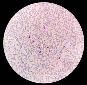
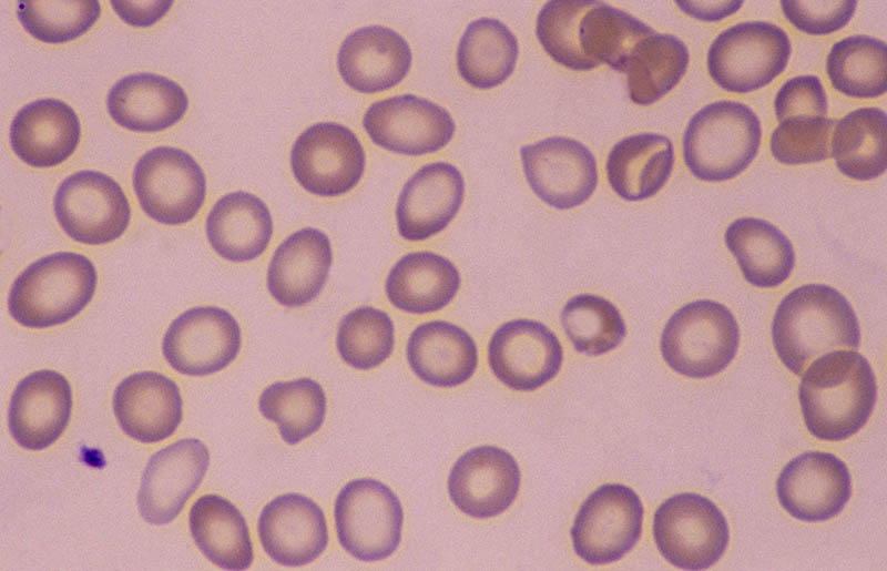
Morphological Abnormalities

Findings:

  • Microcytosis

  • Hypochromia

  • Anisocytosis

  • Poikilocytosis

Diagnostic Clues from Cell Shape

Target cells? Absent
Spherocytes? Absent
Schistocytes? Absent

This makes hemolysis unlikely.

Step 3: Reticulocyte Count and Bone Marrow Response

Reticulocyte count: Low (0.5%)

In severe anemia, we expect increased reticulocytes if bone marrow is responding properly.

Low reticulocytes suggest:

  • Nutritional deficiency

  • Bone marrow suppression

Iron deficiency becomes more likely.

Step 4: Iron Studies Interpretation

TestResultNormal
Serum Ferritin8 ng/mL20–200 ng/mL
Serum IronLowNormal
TIBCHighNormal
Transferrin Saturation10%20–50%

Serum Ferritin

Ferritin is the most specific marker for iron deficiency.
Low ferritin confirms depleted iron stores.

Serum Iron and TIBC

Low serum iron + high TIBC = classic iron deficiency pattern.

Transferrin Saturation

Low transferrin saturation confirms poor iron availability.

Diagnosis strongly supports iron deficiency anemia.

Step 5: Vitamin B12 and Folate Testing

macrocytic aneimia folic acid vitB12 def
macrocytic anemia with thrombocytopenia
hypersegmented neutrophils.2

Vitamin B12: Normal
Folate: Normal

These findings rule out macrocytic anemia causes.

Step 6: Additional Investigations

  • Stool occult blood: Negative

  • Renal function: Normal

  • LDH and bilirubin: Normal

No signs of hemolysis or chronic kidney disease.

Differential Diagnosis of Severe Anemia

  1. Iron deficiency anemia (most likely)

  2. Thalassemia minor

  3. Anemia of chronic disease

  4. Sideroblastic anemia

Lab pattern matches iron deficiency.

Final Diagnosis and Clinical Reasoning

This Severe Anemia Case Study (Step-by-Step Lab Interpretation) demonstrates:

  • Microcytic hypochromic anemia

  • High RDW

  • Low ferritin

  • Low reticulocyte count

  • Heavy menstrual bleeding history

Final Diagnosis: Severe Iron Deficiency Anemia secondary to chronic blood loss.

Treatment Plan and Follow-Up Strategy

Immediate Management

  • Blood transfusion if symptomatic

  • Oral iron therapy (ferrous sulfate 325 mg TID)

Long-Term Plan

  • Investigate menstrual bleeding

  • Dietary counseling

  • Monitor hemoglobin every 4 weeks

Iron therapy typically raises hemoglobin by 1 g/dL per week.

Complications of Untreated Severe Anemia

  • Heart failure

  • Cognitive impairment

  • Pregnancy complications

  • Increased mortality risk

Early diagnosis is critical.

Preventive Strategies

  • Iron-rich diet

  • Routine CBC screening

  • Treat underlying causes

  • Public health awareness

For clinical guidelines, refer to the World Health Organization:
https://www.who.int/health-topics/anaemia

Frequently Asked Questions (FAQs)

1. What is the first test to order in suspected severe anemia?

A Complete Blood Count (CBC).

2. Why is ferritin important?

Ferritin reflects iron stores and confirms deficiency.

3. Can severe anemia be life-threatening?

Yes, especially if hemoglobin falls below 7 g/dL.

4. How quickly does iron therapy work?

Improvement begins within 1–2 weeks.

5. When is transfusion required?

If hemoglobin is critically low with symptoms.

6. What causes microcytic anemia?

Iron deficiency, thalassemia, and chronic disease.

This Severe Anemia Case Study (Step-by-Step Lab Interpretation) illustrates how systematic laboratory interpretation leads to accurate diagnosis. By analyzing CBC indices, peripheral smear findings, reticulocyte count, and iron studies, we identified iron deficiency as the root cause.Understanding this step-by-step method ensures safe clinical decision-making and improved patient outcomes.


Scroll to Top
Review Your Cart
0
Add Coupon Code
Subtotal